
Timisoara_Med 2024, 2023(2), 2; doi:10.35995/tmj20230202
Article
Myotonometric Assessment of the Short-Term Effects of Manual Therapy on Upper Trapezius Myofascial Trigger Points
Iulia Burcos 1,
Andreea Vătăman (Tălîngă) 2
and
Roxana Ramona Onofrei 3,*
1
“Victor Babes” University of Medicine and Pharmacy Timisoara, Physiotherapy Program, 300041 Timisoara, Romania; iuliaburcos@yahoo.com
2
Doctoral School, “Victor Babeş” University of Medicine and Pharmacy Timisoara, 300041 Timisoara, Romania; vataman.andreea@yahoo.com
3
Department of Rehabilitation, Physical Medicine and Rheumatology, Research Center for Assessment of Human Motion, Functionality and Disability, “Victor Babes” University of Medicine and Pharmacy Timisoara, 300041 Timisoara, Romania
*
Correspondence: onofrei.roxana@umft.ro
How to cite: SBurcos, I.; Vătăman (Tălîngă), A.; Onofrei, R.R. Myotonometric Assessment of the Short-Term Effects of Manual Therapy on Upper Trapezius Myofascial Trigger Points. Timisoara Med. 2023, 2023 (Issue 2), 2; doi:10.35995/tmj20230202.
Received: 25 April 2021 / Accepted: 21 December 2023 / Published: 15 January 2024
Abstract
:(1) Background: The aim of this study was to assess the short-term effects of one manual therapeutic intervention by quantifying the biomechanical and viscoelastic properties of upper trapezius myofascial trigger points. (2) Materials and Methods: Fourteen volunteer subjects (six males and eight females) aged 22 to 47 years, with myofascial trigger points in the upper trapezius muscle on both sides participated in this study. The biomechanical and viscoelastic properties were assessed with the Myoton Pro (Estonia) before the application of manual therapy (T0), then immediately after the end of the treatment session (T1) and finally one hour after the end of the treatment session (T2) in order to evaluate the effectiveness of the applied treatment. (3) Results: The oscillation frequency in the right upper trapezius muscle was significantly lower than in the left upper trapezius muscle both immediately, after the end of the manual therapy session (p = 0.03) and one hour after the session (p = 0.03). Regarding the state of tension in the right upper trapezius, a significant decrease was observed one hour after treatment compared to baseline (p = 0.04). In the left upper trapezius muscle, one hour after treatment, the dynamic stiffness value was significantly higher than in the right upper trapezius (p = 0.01). (4) Conclusions: Myotonometry could become a useful method to assess the biomechanical and viscoelastic properties of muscles, identifying trigger points and monitoring the results of therapy.
Keywords:
myotonometry; biomechanical properties; myofascial trigger pointsIntroduction
A myofascial trigger point is defined as a hyperirritable spot found in a taut band in a skeletal muscle, generating local or referred pain and motor disfunction when stimulated by palpation, pressure or movement [1,2]. There are active trigger points, that are associated with local or referred pain, and latent trigger points, that are activated only by different stimuli, like compression, overuse, prolonged stress, poor posture or muscle imbalances [3]. Both types of trigger points have a negative impact on functionality, through their effects on muscle activation and relation pattern, range of motion or muscle stiffness. Trigger points in the upper trapezius muscle are common [4], as a result of muscle overloading or overuse due to prolonged static posture, for example, in sedentary sitting jobs.
There are several proposed treatments that have been proved to be efficient, like ischemic compression, muscle energy techniques, different physical therapy procedures or dry needling [3]. The efficacy of these treatments has been assessed mainly by patient-reported outcomes. There are studies that have also used the assessment of muscle biomechanical properties to evaluate trigger point treatments [5,6,7].
There is no gold standard for the assessment and diagnosis of trigger points. The diagnosis is based on the identification of the trigger point in a taut band, with symptom reproduction [2], a maneuver dependent on the examiner’s skills and experience. Other assessment and diagnostic techniques have been proposed, like ultrasound elastography [8] or myotonometry, based on the viscoelastic and biomechanical properties of trigger points. Myotonometry has been shown to be a reliable tool that was able to quantify myofascial tissues’ viscoelastic properties [9]. Myoton Pro is a hand-held device that assesses muscles’ viscoelastic and biomechanical properties in a simple, non-invasive and reliable manner [10]. Several studies have used myotonometry to assess the change in these properties at the level of trigger points as a measure of treatment outcomes [6,7,9].
The aim of this study was to assess the short-term effects of manual therapy through the quantification of the biomechanical and viscoelastic properties of the upper trapezius myofascial trigger points. We hypothesized that manual therapy would produce a rapid decrease in tension and stiffness, with an increase in elasticity at the level of trigger points.
Materials and Methods
Fourteen volunteer consecutive subjects with myofascial trigger points in the upper trapezius muscles on both sides participated in this observational, prospective study. The inclusion criteria were: (a) aged over 18 years; (b) presence of myofascial trigger points in the upper trapezius; (c) no history of trauma, injuries or surgeries at the level of the neck and shoulder girdle. The study was carried out in accordance with the Declaration of Helsinki and was approved by the institutional Ethical Committee (no 25/28.03.2022)
The presence of the myofascial trigger points was confirmed during clinical examination—palpation and compression of the trigger points reproduced the localized or referred pain and were marked with a pen. For testing, the subjects had to be relaxed. They were advised not to perform strenuous physical activity the day before testing.
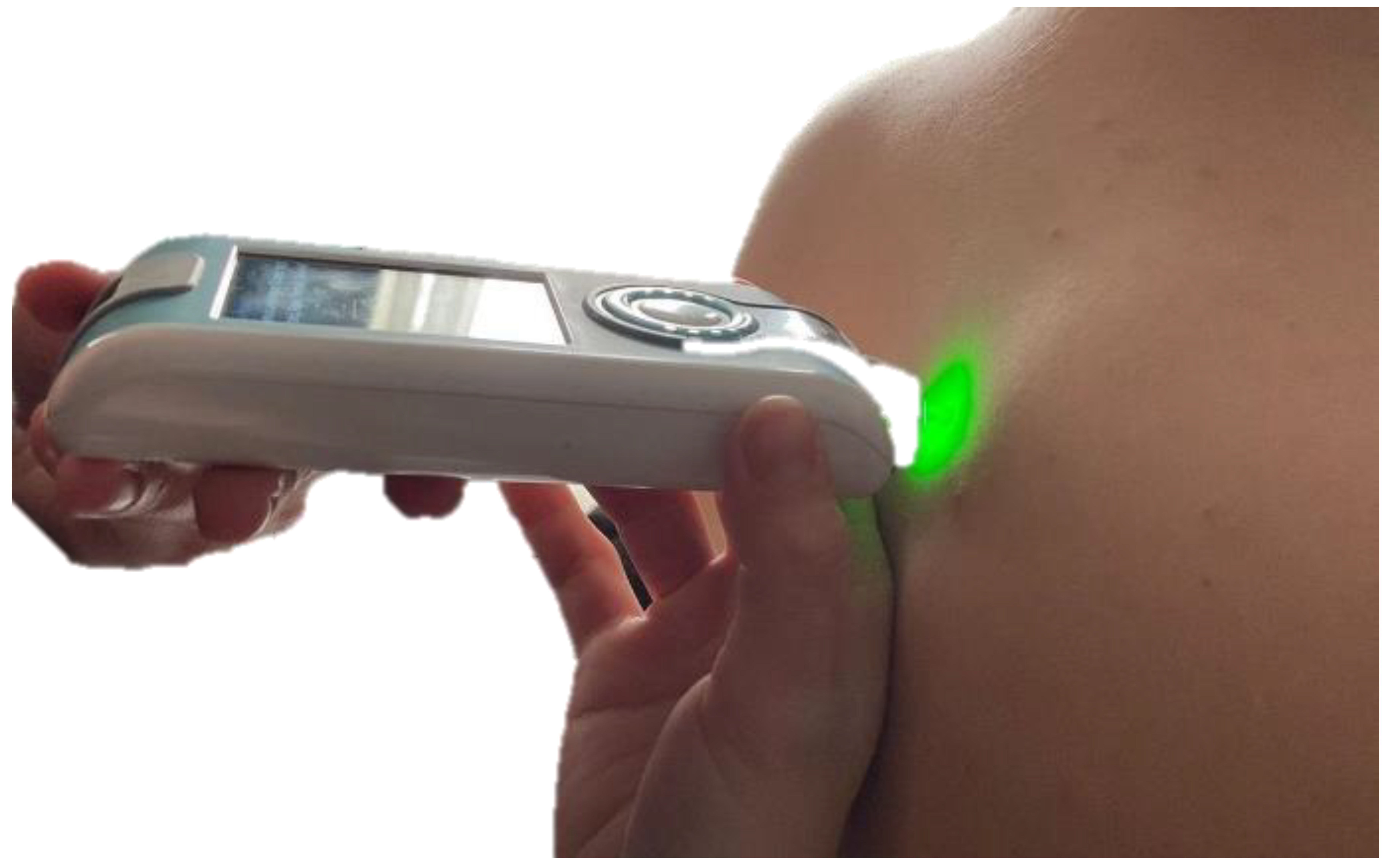
The biomechanical and viscoelastic properties were assessed with a hand-held myotonometer, the Myoton Pro (Estonia) [11] (The device was placed over the trigger points and provided 5 pulses at 0.8 second intervals; the results displayed were the arithmetic averages of these determinations. The device was positioned perpendicular to the test point and held in this position throughout the evaluation (Figure 1). The results were saved in the device and then transferred to the corresponding program. Clinical examination and myotonometric testing were performed by the same researcher, and the data were analyzed by a different researcher.
The evaluated parameters were oscillation frequency, dynamic stiffness, logarithmic decrement, relaxation time and creep. The oscillation frequency (Hz) indicates the state of tension at the level of the tested point; dynamic stiffness (N/m) represents the resistance of the muscle to the forces acting on it and tending to deform it; logarithmic decrement provides information on the tissue elasticity; relaxation time (ms) represents the time taken by the muscle to return to its original shape and creep is the ratio between relaxation time and deformation time of the muscle [11] Trigger points were assessed bilaterally at the level of the upper trapezius muscles.
Figure 1.
Myotonometric assessment.
The assessments were performed before manual therapy (T0), immediately after the end of the treatment session (T1) and one hour after the end of the treatment session (T2) in order to evaluate the effectiveness of the applied treatment. The therapy consisted mainly of the ischemic compression method applied at the level of upper trapezius trigger points. The pressure was maintained for 5 seconds, with a 3 second pause.
Statistical analysis was performed with the MedCalc software version 22.009 (Ostend, Belgium). Data are presented as means and standard deviations. The repeated measures ANOVA with Bonferroni post-hoc test was used to analyze differences between ratings for each individual parameter and muscle. The paired Student’s t-test was used for comparisons between the values obtained between the muscle on the right and left side. Differences were considered significant for values of p < 0.05.
Results
The study included 14 subjects aged between 22 and 47 years, involved in static occupational activities. The mean age was 31.5 ± 8.75 years; all subjects were of normal weight (BMI—23.51 ± 3.18 kg/m2; weight 70 ± 13.47 kg; height 172.79 ± 11.13 cm). Of the subjects included in the study group, six were male (42.86%) and eight female (57.14%). All subjects had the right hand as the dominant hand. Subjects’ characteristics are presented in Table 1.

Table 1.
Subjects’ characteristics.
| Subjects (n = 14) | |
|---|---|
| Age (years), mean ± SD | 31.5 ± 8.75 |
| Weight (kg), mean ± SD | 70 ± 13.47 |
| Height (cm), mean ± SD | 172.79 ± 11.13 |
| BMI (kg/m2), mean ± SD | 23.51 ± 3.18 |
All biomechanical and viscoelastic parameter measured before and after treatment are presented in Table 2.
Table 2.
Biomechanical and viscoelastic parameters measured at the level of upper trapezius trigger points, before and after treatment.
Table 2.
Biomechanical and viscoelastic parameters measured at the level of upper trapezius trigger points, before and after treatment.
| T0 | T1 | T2 | ||||
|---|---|---|---|---|---|---|
| Right | Left | Right | Left | Right | Left | |
| Oscillation frequency (Hz) | 17.62 ± 2.47 | 17.66 ± 2.64 | 16.78 ± 2.4 | 17.64 ± 2.55 | 16.44 ± 2.29 | 17.21 ± 1.71 |
| Dynamic stiffness (N/m) | 341.07 ± 64.81 | 341.5 ± 73.04 | 327.78 ± 74.11 | 336.85 ± 66.34 | 307.57 ± 64.18 | 334 ± 40.33 |
| Logarithmic decrement | 1.34 ± 0.34 | 1.39 ± 0.31 | 1.37 ± 0.35 | 1.24 ± 0.27 | 1.31 ± 0.34 | 1.3 ± 0.29 |
| Relaxation time (ms) | 15.82 ± 2.95 | 15.99 ± 3.3 | 16.66 ± 3.19 | 16.05 ± 3.44 | 17.21 ± 1.71 | 16.02 ± 1.86 |
| Creep | 0.97 ± 0.16 | 0.99 ± 0.18 | 1.02 ± 0.17 | 0.99 ± 0.19 | 1.06 ± 0.13 | 0.99 ± 0.11 |
Data are presented as mean ± SD.
The oscillation frequency in the right upper trapezius muscle was significantly lower in T2, at one hour after the manual therapy session, compared to T0 (p = 0.04). Although there was a decrease in the dynamic stiffness, the differences between assessments were not statistically significant. Although an improvement in elasticity was observed immediately after the end of the therapy session in the right upper trapezius, the differences were not statistically significant, and this improvement in elasticity was not maintained one hour after treatment. No improvement in relaxation time required for the muscle to return to its original shape was observed immediately after the treatment session. The creep parameter, representing the ratio between relaxation time to deformation time, did not show significant changes after treatment.
At baseline, no side-to-side significant differences were found for none of the assessed parameters. At T1 and T2, the oscillation frequency was significantly lower in the right upper trapezius compared to left side (p = 0.03 for both comparisons).
In the left upper trapezius muscle, at T2, the dynamic stiffness value was significantly higher than in the right upper trapezius (p = 0.01), while the relaxation time one hour after treatment (T2) was significantly lower than in the right upper trapezius (p = 0.01).
Discussion
The biomechanical and viscoelastic properties of the upper trapezius myofascial trigger points were assessed using myotonometry to evaluate whether there are short-term changes after applying a manual therapeutic intervention. Our hypothesis that a manual therapeutic intervention would produce a rapid decrease in tension and stiffness, with an increase in elasticity at the level of trigger points, was partially confirmed. Our results showed that only tension, expressed by the oscillation frequency parameter, decreased significantly at one hour after treatment and only on the right upper trapezius trigger points. Stiffness also showed lower values after treatment; however, the observed changes were not statistically significant.
Similar results, with a decrease in tension and stiffness immediately after the first therapeutic session, were reported by Olesiejuk et al. [6]. They applied the ischemic compression method of myofascial trigger points, located in the upper trapezius muscle, to a group of thirty-one female patients with episodic migraine. The immediate effects reported by Olesiejuk et al. were observed only in the left upper trapezius. Their explanation for the immediate unilateral decrease was due to the fact that the tested subjects were right-handed, using their right side more frequently in daily life, so the higher hypertonia in the right upper trapezius did not permit a significant decrease after only one therapeutic session. However, the subjects assessed in our study were also right-handed, and we observed a significant decrease in tension only on the right side. Moreover, we found significantly lower values in tension and stiffness on the right upper trapezius compared to the left side both immediately and at one hour after the therapeutic session, although before treatment the values were not statistically different.
We observed a decrease in stiffness, similar to Wendt et al. [12] who also noticed an immediate effect on muscle tone and stiffness immediately after applying different therapeutic procedures for the treatment of upper trapezius trigger points, mainly on the right side. Kisilewicz et al. [5] also reported a decrease in stiffness in the upper trapezius after one session of ischemic compression treatment in a group of basketball players.
As reported in other studies, myotonometry proved to be a sensible method to assess differences in muscle viscoelastic and biomechanical parameters as a result of different treatments. The results of Jimenez-Sanchez et al. [9] support the ability of the myotonometer to identify and detect differences between the mechanical properties of myofascial trigger points. Perez-Bellmunt et al. [13] also reported a decrease in stiffness in latent trigger points in gastrocnemius muscles after a single therapeutic session.
The limitations of the present study should be noted, with the relatively small sample size being one limitation. Another limitation could be the subjectivity of trigger point identification conducted by only one researcher. Further studies on larger sample size, including subjects with both latent and active myofascial trigger points, as well as assessments after longer treatments using different therapeutic approaches are needed.
Conclusions
The results of this study showed a significant decrease in mechanical tension one hour after the end of the session in the right upper trapezius muscle. Analysis of changes in the biomechanical and visco-elastic properties at trigger points can provide useful information for understanding pathogenic mechanisms, but also for adjusting therapies. Myotonometry could be a valuable method of assessment, due to its portability, relatively low cost and its ease of use.
Author Contributions
Conceptualization, R.R.O. and I.B.; methodology, R.R.O.; validation, R.R.O., I.B. and A.V.; formal analysis, I.B. and R.R.O.; investigation, I.B.; data curation, R.R.O., I.B. and A.V.; writing—original draft preparation, I.B., A.V. and R.R.O.; writing—review and editing, I.B., A.V. and R.R.O.; visualization, R.R.O.; supervision, R.R.O. All authors have read and agreed to the published version of the manuscript.
Funding
This research received no external funding.
Conflicts of Interest
The authors declare no conflicts of interest.
References
- Simons, D.G. Review of Enigmatic MTrPs as a Common Cause of Enigmatic Musculoskeletal Pain and Dysfunction. J. Electromyogr. Kinesiol. 2004, 14, 95–107. [Google Scholar] [CrossRef] [PubMed]
- Travell, J.G.; Simons, D.G. Myofascial Pain and Dysfunction: The Trigger Point Manual; Lippincott Williams & Wilkins: Philadelphia, PA, USA, 1992; Volume 2, ISBN 0683083678. [Google Scholar]
- Huguenin, L.K. Myofascial Trigger Points: The Current Evidence. Phys. Ther. Sport 2004, 5, 2–12. [Google Scholar] [CrossRef]
- Chang, C.-W.; Chang, K.-Y.; Chen, Y.-R.; Kuo, P.-L. Electrophysiologic Evidence of Spinal Accessory Neuropathy in Patients With Cervical Myofascial Pain Syndrome. Arch. Phys. Med. Rehabil. 2011, 92, 935–940. [Google Scholar] [CrossRef] [PubMed]
- Kisilewicz, A.; Janusiak, M.; Szafraniec, R.; Smoter, M.; Ciszek, B.; Madeleine, P.; Fernández-de-Las-Peñas, C.; Kawczyński, A. Changes in Muscle Stiffness of the Trapezius Muscle after Application of Ischemic Compression into Myofascial Trigger Points in Professional Basketball Players. J. Hum. Kinet. 2018, 64, 35. [Google Scholar] [CrossRef] [PubMed]
- Olesiejuk, M.; Marusiak, J.; Chalimoniuk, M. Myofascial Trigger Points Therapy Decreases Myotonometric Tone and Stiffness of Trapezius Muscle, Benefits Headaches and Muscle Pain in Migraine. NeuroRehabilitation 2023, 52, 299–310. [Google Scholar] [CrossRef] [PubMed]
- Jiménez-Sánchez, C.; Gómez-Soriano, J.; Bravo-Esteban, E.; Mayoral-del Moral, O.; Herrero-Gállego, P.; Serrano-Muñoz, D.; Ortiz-Lucas, M. Effects of Dry Needling on Biomechanical Properties of the Myofascial Trigger Points Measured by Myotonometry: A Randomized Controlled Trial. J. Manip. Physiol. Ther. 2021, 44, 467–474. [Google Scholar] [CrossRef] [PubMed]
- Sikdar, S.; Shah, J.P.; Gilliams, E.; Gebreab, T.; Gerber, L.H. Assessment of Myofascial Trigger Points (MTrPs): A New Application of Ultrasound Imaging and Vibration Sonoelastography. In Proceedings of the 2008 30th Annual International Conference of the IEEE Engineering in Medicine and Biology Society, Vancouver, BC, Canada, 20–25 August 2008; IEEE: New York, NY, USA, 2008; pp. 5585–5588. [Google Scholar]
- Jiménez-Sánchez, C.; Ortiz-Lucas, M.; Bravo-Esteban, E.; Mayoral-del Moral, O.; Herrero-Gállego, P.; Gómez-Soriano, J. Myotonometry as a Measure to Detect Myofascial Trigger Points: An Inter-Rater Reliability Study. Physiol. Meas. 2018, 39, 115004. [Google Scholar] [CrossRef] [PubMed]
- Taş, S.; Yaşar, Ü.; Kaynak, B.A. Interrater and Intrarater Reliability of a Handheld Myotonometer in Measuring Mechanical Properties of the Neck and Orofacial Muscles. J. Manip. Physiol. Ther. 2021, 44, 42–48. [Google Scholar] [CrossRef] [PubMed]
- Available online: https://www.myoton.com (accessed on 1 February 2023).
- Wendt, M.; Kocur, P.; Lewandowski, J.; Waszak, M. Effect of the Combined Therapy of the Muscle Energy Technique and Trigger Point Therapy on the Biophysical Parameters of the Trapezius Muscle: A Randomized Clinical Trial. Muscles Ligaments Tendons J. (MLTJ) 2021, 11, 41. [Google Scholar] [CrossRef]
- Pérez-Bellmunt, A.; Simon, M.; López-de-Celis, C.; Ortiz-Miguel, S.; González-Rueda, V.; Fernandez-de-las-Peñas, C. Effects on Neuromuscular Function After Ischemic Compression in Latent Trigger Points in the Gastrocnemius Muscles: A Randomized Within-Participant Clinical Trial. J. Manip. Physiol. Ther. 2022, 45, 490–496. [Google Scholar] [CrossRef] [PubMed]
© 2024 Copyright by the authors. Licensed as an open access article using a CC BY 4.0 license.